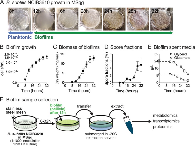
FIG 1.
Biofilm growth and sample handling. (A) B. subtilis NCIB3610 was inoculated into modified MSgg media at time t = 0. Standing cultures at 8 h after inoculation were planktonic. Initial pellicle formation (i.e., fragile biofilms) reproducibly took place at approximately 12 h. By 16 h, pellicles became noticeably more robust and the wrinkled morphology started to become apparent. Biofilm development was monitored until 32 h of growth. The pictures shown are representative of all experiments performed. (B) Biofilm growth was quantified in cells per milliliter (direct cell counts). Data represent averages of results from 4 to 6 biological replicates ± standard errors of the means (SEM). (C) Biomass of B. subtilis biofilms. Extracted pellicles were air-dried and weighed. Data represent averages of results from 3 biological replicates ± SEM. (D) Spore fractions (percent) of biofilms. Data represent averages of results from 5 biological replicates ± SEM. (E) Glycerol and glutamate concentrations in spent biofilm media over the course of biofilm growth. Data represent averages of results from 3 biological replicates ± SEM. (F) Sample collection for metabolomic, transcriptomic, and proteomic analyses was performed at 4-h intervals from 8 to 32 h of growth at 37°C. Planktonic cells (8 h) were collected via rapid filtration. Biofilm samples (12 to 32 h) were collected by lifting a custom-made stainless-steel mesh and transferring the pellicle to the appropriate extraction solvent or buffer.

