
Figure 2.
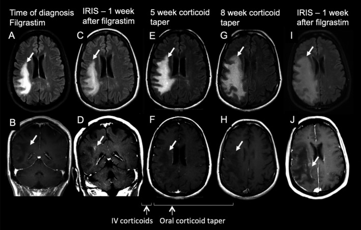
Progression of PML lesions after premature corticosteroid treatment. Axial FLAIR (A, C, E, G, I) and postcontrast T1‐weighted images (B, D, F, H, J) demonstrate PML lesion in the right frontoparietal area (arrows) of one representative MS patient. A, B) The PML lesion is devoid of contrast enhancement at the time of PML diagnosis. (C, D) Contrast enhancement on MRI and clinical worsening indicative of IRIS occurred 9 days after peak ALC on filgrastim treatment and 63 days after Nz withdrawal. The patient received intravenous methylprednisolone followed by oral prednisone taper. Three to 4 weeks later, while on prednisone taper, the patient began to worsen clinically. Repeat brain MRI exams revealed increased size PML lesions without contrast enhancement (E, F, G, and H). Prednisone was discontinued and filgrastim reinstituted. There was evidence of a more robust IRIS by contrast enhancement on MRI 1 week after the second filgrastim treatment (I, J).