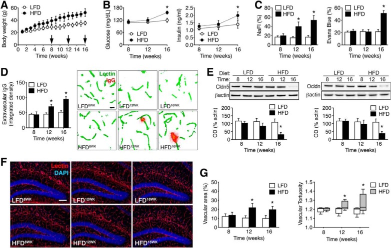
Figure 1.
Progressive changes in BBB integrity with increasing durations of obesogenic diet consumption. A, Weight gain with exposure to a HFD or LFD in male mice. B, Fasting hyperglycemia (left) and fasting hyperinsulinemia (right) were evident after 16 weeks consumption of HFD. C, Increased penetration of the low molecular weight tracer NaFl was observed in clarified hippocampal lysates after 12 or 16 weeks HFD (left graph). Right graph shows increases in hippocampal Evans blue after 16 weeks HFD. D, Accumulation of extravascular IgG deposits occurs in the dentate molecular layer after 12 and 16 weeks HFD. Micrographs (right) show extravascular IgG immunoreactivity after background subtraction, as described previously (Bell et al., 2010). Scale bar: (in LFD8WK) for all panels, 10 μm. E, Reductions in hippocampal claudin-5 (left) and occludin (right) protein were detected by Western blotting after 16 weeks of HFD. F, Micrographs show representative images of tomato lectin labeling for the indicated diets and time points. Scale bar: (in LFD12WK) for all micrographs, 100 μm. G, Increases in vascular area (left) and tortuosity (right) in the dentate molecular layer with increasing durations of HFD. For all graphs, bar or symbol height represents the mean of (n = 4–6) mice per diet at each time point and error bars represent SEM. *Indicates significant differences (p < 0.05) determined by ANOVA.

