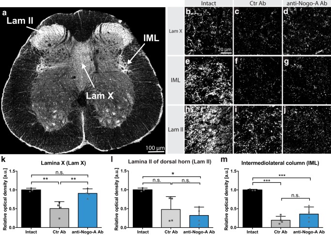
Figure 5.
Innervation of lumbosacral spinal cord by CRF-positive fibers, including bulbospinal fibers from the PMC at 28 d after injury. a, In intact rats, immunofluorescent CRF-positive fibers and terminals are concentrated in dorsal horn Lam II, the IML, and Lam X. b–j, High-magnification image (40×) of Lamina X (b–d), IML column (e–g), and Lam II (h–j) in intact, spinal cord-injured control antibody-treated (Ctr Ab), and injured anti-Nogo-A antibody-treated rats. k–m, Relative optical density quantitation for CRF in Lam X, IML, and Lam II. Data are presented as means ± SD. Scale bars: a, 100 μm; b–j, 20 μm; ns, not significant; *p < 0.05; **p < 0.01; ***p < 0.001.

