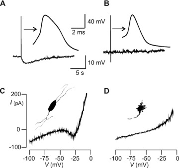
Figure 1.

Identification of myenteric neurons in rat colon. (A & C) AH cell and (B & D) S cell. (A) AH and (B) S cell membrane voltage recording. Arrows point to action potentials on expanded time base. Hump during action potential repolarization and sAHP > 2 sec. duration after single AP distinguishes AH from S cell. (C) Non‐monotonic quasi‐steady‐state I–V curve from AH cell. Inset portrays neuron silhouette showing multipolar Dogiel type II morphotype. (D) I–V trace from S cell is monotonic and lacks region of negative conductance. Inset shows uniaxonal cell silhouette with multiple short dendrites indicating the Dogiel type I morphotype.
