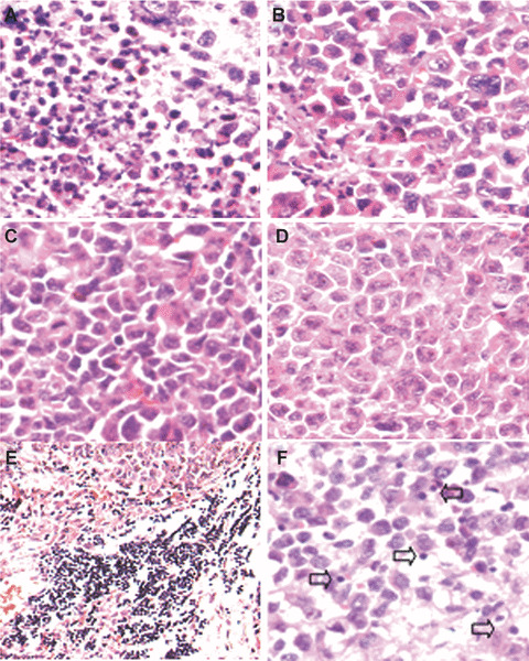
Figure 3.

Histological analysis of tumour tissue. Histological examination of tumours by haematoxylin and eosin staining was performed. Images show paraffin‐embedded tumour sections from tumour‐bearing mice treated with p‐c (A), e‐p (B), liposome alone (C) or PBS (D). There were large areas of confluent tumour cells with little or no tumour tissue necrosis (B–D) but extensive necrosis (A), respectively. Meanwhile, lymphocyte infiltration was also enhanced both in the margin (E) and central regions in p‐c treated group (F).
