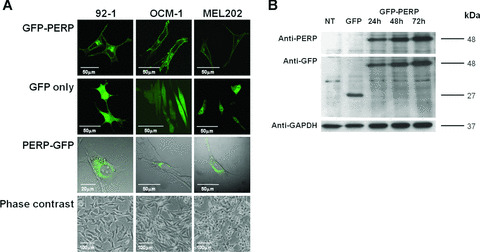
Figure 1.

(A) Confocal images showing transiently transfected living 92‐1, OCM‐1 and MEL202 cells expressing GFP‐PERP, GFP only and PERP‐GFP at 24 hr after transfection. GFP‐PERP fusion protein is targeted to the plasma membrane, although the presence of the fluorescent tag at the C‐terminus of PERP impairs this targeting (bright field is shown for pPERP‐GFP transfected cells to illustrate the lack of plasma membrane targeting of the PERP‐GFP fusion protein). Phase‐contrast images illustrate the characteristic cell types of the cell lines used, specifically, 92‐1 cells being predominantly epithelioid, OCM‐1 being predominantly spindle cell type and MEL202 being mixed (epithelioid and spindle) cell type, which are features that assist in the prediction of metastatic disease. (B) Lysates of cells transfected with pGFP‐PERP were analysed at 24, 48 and 72 hrs after transfection by Western blotting using anti‐PERP and anti‐GFP antibody. Nontransfected (NT) and pEGFP‐N1 (GFP only)‐transfected cells served as controls. GFP‐PERP fusion protein of the expected 48 kD size was detected by both antibodies in pGFP‐PERP‐transfected cells only, whereas the 27 kD GFP protein was detected only by the anti‐GFP antibody in pEGFP‐N1‐transfected cells. The same blot was sequentially probed with anti‐GAPDH antibody to confirm equal loading. GFP‐PERP fusion protein was identically detected in all UM cells studied (blot shown is of MEL202 cell lysates).
