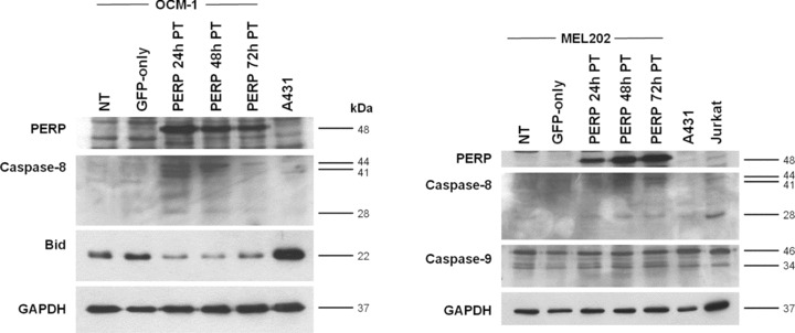
Figure 4.

Caspase activation following PERP expression. Cell lysates were prepared from pGFP‐PERP‐transfected UM cells at 24, 48 and 72 hrs after transfection. Lysates from nontransfected (NT) and pEGFP‐N1‐transfected (GFP‐only) cells were harvested 48 hrs after transfection and served as controls. A431 and Jurkat cell lysates served as positive controls for the specified antibodies, as recommended by the manufacturers of the respective antibodies. Lysates were analysed by Western blotting for the processing of initiator caspase‐8 and ‐9 and the cleavage of Bid. Molecular weights of detected cleaved caspase proteins and full‐length Bid are indicated. Images referring to the same cell line are of the same blot probed sequentially with the specified antibodies. Detection of GAPDH was used to confirm equal loading of samples. Partially processed caspase‐8 proteins (44, 41 and 28 kD) were readily detected in UM cells expressing GFP‐PERP. In addition, full‐length Bid levels were depleted in cells expressing GFP‐PERP compared with control cells. With regard to caspase‐9, there was no difference in the levels of cleaved enzyme (34 kD) in cells expressing GFP‐PERP, GFP only, or nontransfected cells.
