
Figure 5.
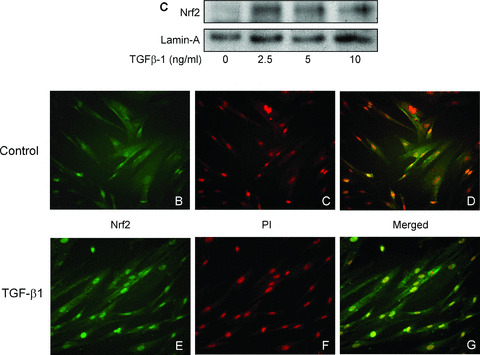
Nuclear translocation of Nrf2 in HAoSMC treated with TGF‐β1. Confluent cultures were equilibrated in medium containing 1% FCS prior to treatment with TGF‐β1 (2.5–10 ng/ml, 2 hrs). (A) Immunoblot analysis of Nrf2 protein expression in nuclear extracts relative to the loading control lamin‐A. Cellular localization of Nrf2 was determined by immunofluorescent microscopy in untreated cells (B–D) and cells treated with TGF‐β1 (2.5 ng/ml, 2 hrs, E–G) and stained using a specific antibody against Nrf2 and an Alexa Fluor‐488 conjugated secondary antibody (B, E). Nuclei were counterstained with propidium iodide (C, F) and images merged using Photoshop software (D, G). Data are representative of similar results in three different cell cultures.