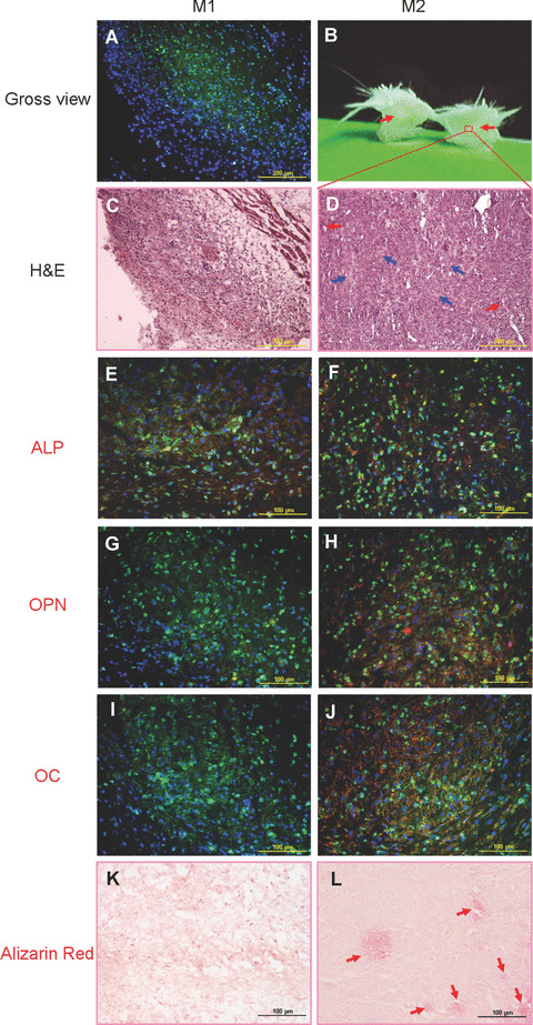
Figure 7.

In vivo osteogenic differentiation of M1P14 and M2P14. (A) Twelve days after subcutaneous injection of M1P14, only a small area of abdominal skin specimens were stained with MHC‐Ib antibody (green) under microscopic observation (10×), which showed the existence of cell grafts. (B) After cutting across the protuberance of abdominal skin injected with M2P14, a white area (as pointed by red arrows) could be seen in the skin layers subcutaneously. (C) Haematoxylin and eosin staining view of the M1P14 graft specimens (10×). (D) Haematoxylin and eosin staining view of the white area of M2P14 graft specimens (10×). Blood vessel examples filled with red blood cells are pointed by red arrows. And some matrix‐rich areas are indicated by blue arrows. (E) ALP staining (red) could be detected in M1P14 cell graft area (green) (20×). (F) ALP staining (red) could be detected in M2P14 cell graft area (green) (20×). (G) No OPN staining could be detected in M1P14 cell graft area (green) (20×). (H) OPN staining (red) could be detected in M2P14 cell graft area (green) (20×). (I) No OC staining could be detected in M1P14 cell graft area (green) (20×). (J) OC staining (red) could be detected in M2P14 cell graft area (green) (20×). (K) Alizarin red staining showed absence of mineral deposition in M1P14 graft specimens (20×). (L) Alizarin red staining showed some positively stained areas of mineral deposition (as indicated by red arrows) in M2P14 graft specimens (20×). Cell nuclei in all pictures were stained with DAPI (blue).
