Figure 5.

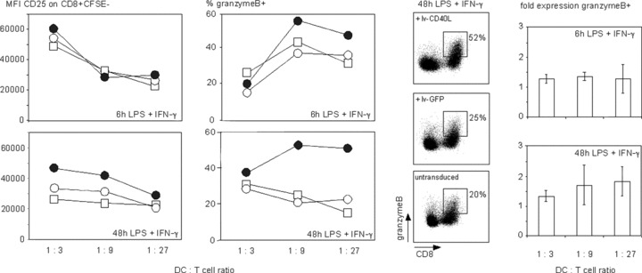
CD40L ectopically expressed on cytokine‐exhausted TLR4‐activated DCs triggers activation of CD8+ T lymphocytes. Allogeneic MLR using DCs that were matured for 6 or 48 hrs upon receiving a 6‐hr LPS/IFN‐γ stimulus as indicated (bottom right of each panel) followed by a 6‐hr treatment with the lenti‐virus lv‐CD40L (black circles) or lv‐GFP (white circles) in comparison to only primary stimulated DCs (squares). T‐cell activation was measured by the mean fluorescence intensity (MFI) of CD25 expressed on proliferating CFSE negative CD8+ T cells. Granzyme B expressing cells are given as the percentage in CD8+ T cells. The dot plots show CD8+ granzyme B+ T cells at a DC:T cell ratio = 1:9. Shown is one representative experiment of three using three different donors. The bar diagram combines the data of three different donors, which shows fold expression ± S.D. of granzyme B expressing CD8+ T cells stimulated with lv‐CD40L DCs normalized to lv‐GFP DCs; 6 hrs LPS/IFN‐γ pre‐matured DCs; P= 0,01; 48 hrs LPS/IFN‐γ pre‐matured DCs, P= 0,004. CD40L or GFP expression as well as the phenotype of the DCs used for the depicted alloMLRs is shown in Fig. 4.
