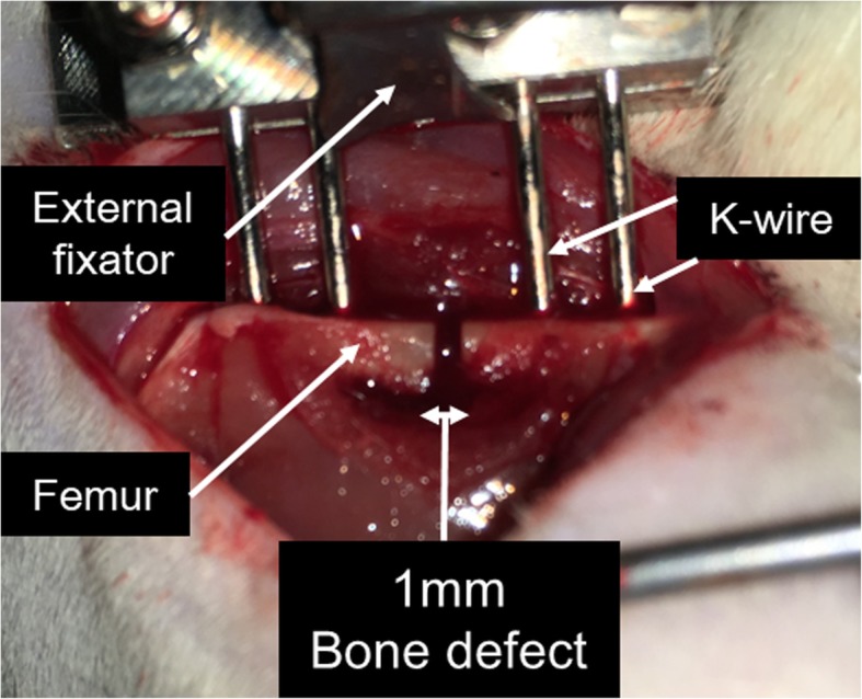
Fig. 1.

Representative intraoperative image. After exposing the femoral shaft, two pairs of tip-threaded 1.4-mm-diameter K-wires were placed in the proximal and distal femur. A bone defect of 1 mm was created between the proximal and distal pins by an oscillating saw, and a custom-made external fixator was attached to connect the pairs of pins
