Fig. 3.

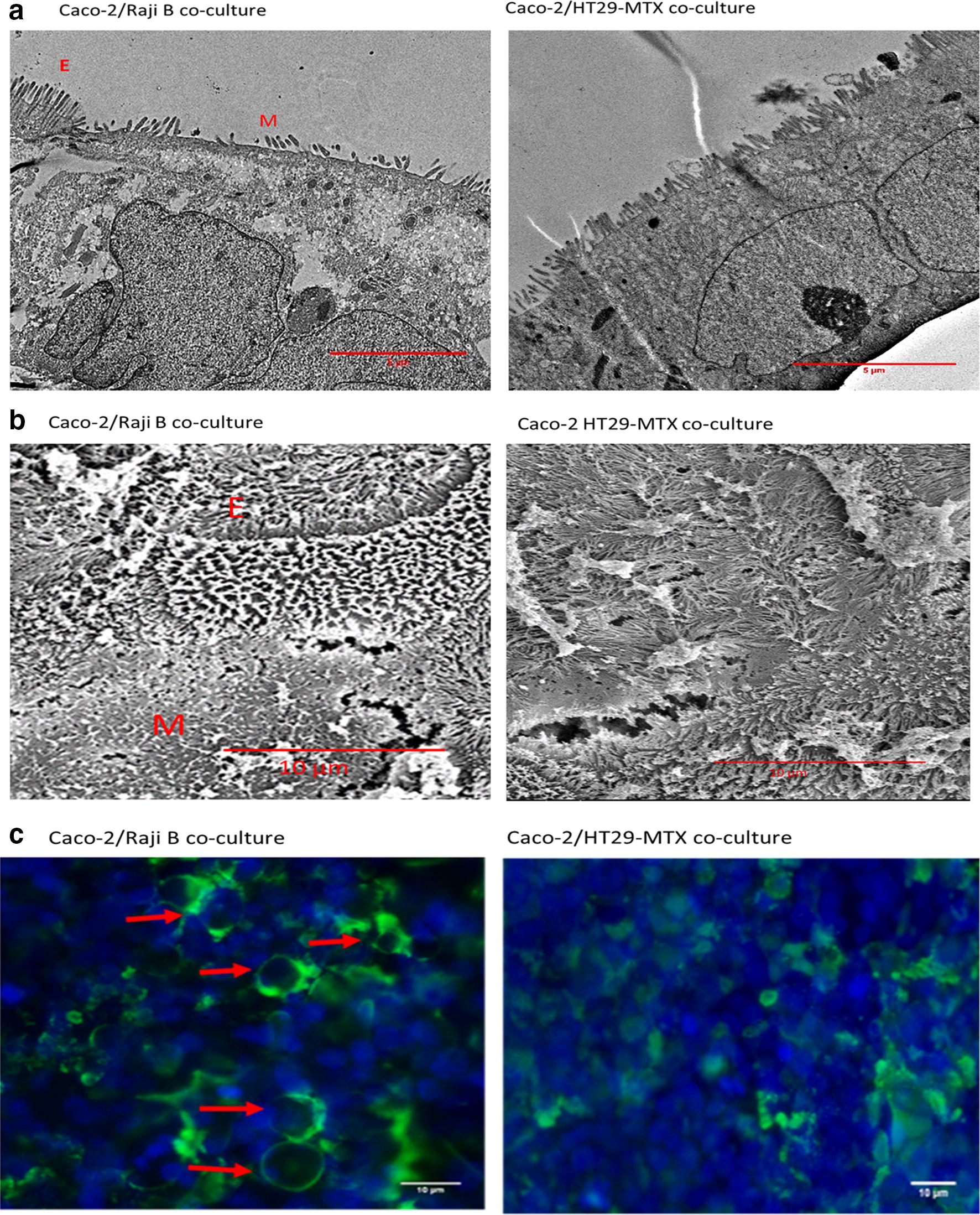
Confirmation of the presence of M cells in Caco-2/Raji B co-culture. To identify the presence of M cells Caco-2/Raji B and Caco-2/HT29-MTX co-cultures were examined by TEM (a) and SEM (b). ‘M’ indicates the presence of M cells, which have a reduced number of microvilli, and ‘E’ indicates epithelial cells with microvilli which cover the cell surface. M cells were also identified using fluorescent microscopy; Caco-2/Raji B and Caco-2/HT29-MTX co-culture models were fixed, labelled with WGA FITC (green) and mounted with ProLong with DAPI (nucleus: blue) and the images obtained with a fluorescent microscope (b). Red arrows indicate the presence of M cells. TEM and SEM scale bar = 5 µm, WGA staining and SEM scale bar = 10 µm. Representative images are presented
