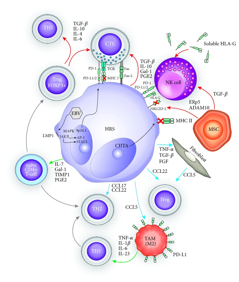
Figure 1.
Immune-escape mechanisms of cHL TME. Chemokine secretion by HRS plays a central role in the TME immunosuppressive state of cHL. They allow differentiation of infiltrating naïve CD4+ T-cells into regulatory FOXP3+ and Th2 T-cells and provide CTL inhibitory signals through TGF-ß, IL-10, galectin-1 (Gal-1), tissue inhibitor of metalloproteinase 1 (TIMP1), and prostaglandin E2 (PGE2). HRS also attract Tregs/Th2 T-cells from the systemic circulation, through the secretion of CCL22 and CCL17, respectively, while at the same time promoting their expansion through the secretion of Gal-1, TIMP1, and PGE2. Fibroblasts also contribute to Treg chemoattraction through CCL5 secretion. In the same line, TAM promote the differentiation of Th1 cells towards the Th2 phenotype. Nevertheless, recent evidence challenges this concept of predominant Th2 polarized TME and evidenced an increase in activated Th1 T-cells in TME of cHL patients. EBV latent infection also plays a key role, through the production of LMP1 which activates the MAPK and JAK/STAT3 pathways leading to transcriptional activation of the 9p24.1 locus with consequent PD-L1/2 overexpression [5]. Even in EBV negative cHL, PD-L1 (and, to a lesser degree, PD-L2) expression at the surface of HRS cells is still high. PD-1-PD-L1 interaction triggers the inhibition of CTL function. TAM display a high surface PD-L1 expression, thus promoting PD-1-PD-L1 axis immune escape. EBV- cHL displays also a decrease in MHC class I expression, in comparison to their EBV positive counterparts mainly through B2-microglobulin subunit downregulation. MHC class II expression can also be impaired through epigenetic silencing, in the subset of mutated/translocated CIITA cHL. In this context, it is presumed that the PD-1/PD-L1/2 axis mediates immune escape, in first instance, by dampening NK cell activity. NK function is further downregulated by TGF-ß secreted by HRS and mesenchymal MSC. MSC also edit the surface expression of NKG2D-L through the enzymatic activity of secreted ERp5 and ADAM10. HRS cells also express Fas ligand at their surface, thus promoting apoptosis of interacting Th1 and cytotoxic T-cells [88]. Red arrows: “inhibition signal”; green arrows: “differentiation signal”; blue arrows: “chemoattraction” signal.

