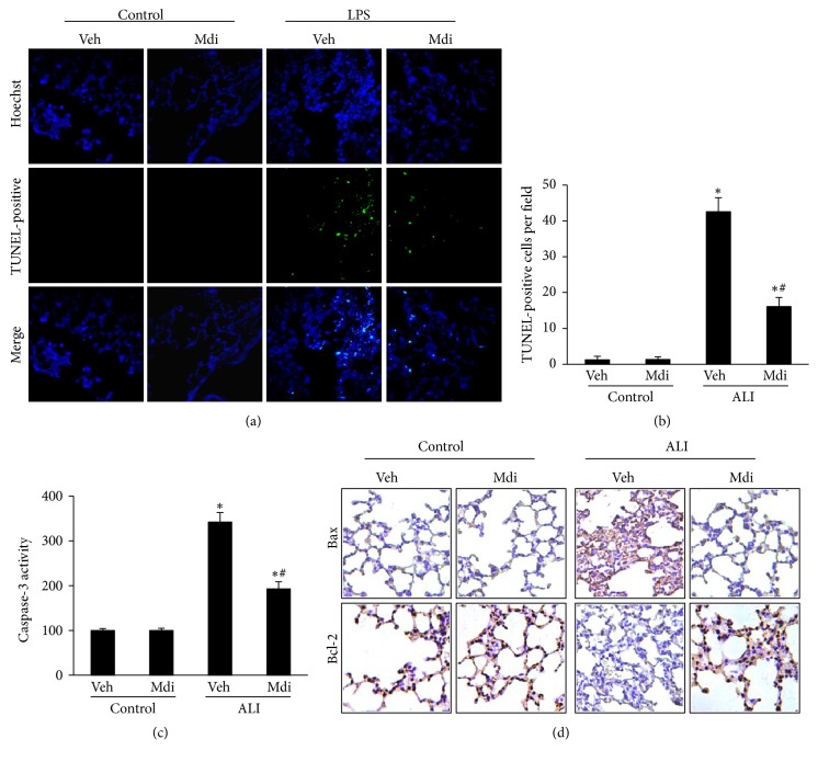
Figure 4.
Mdivi-1 prevents lung cell apoptosis in vivo. (a) The level of pulmonary cell apoptosis increased following LPS administration, as determined by TUNEL staining (200× magnification). (b) TUNEL-positive cells were averaged over 10 microscopic fields per animal. LPS-challenged animals exhibited a significant increase in TUNEL-positive cells, which was reduced by treatment with Mdivi-1 treatment. (c) Caspase-3 activity in the lung tissues was elevated in the LPS group but suppressed in the group treated with Mdivi-1. (d) The level of Bax and Bcl-2 protein expression was determined by immunohistochemistry. The increased Bax expression and decreased Bcl-2 expression in the LPS group was reversed following Mdivi-1. Data are presented as the mean ± SD (n = 6 in each group). ∗P < 0.05 versus control group; #P < 0.05 versus LPS + vehicle group.

