
FIGURE 5.
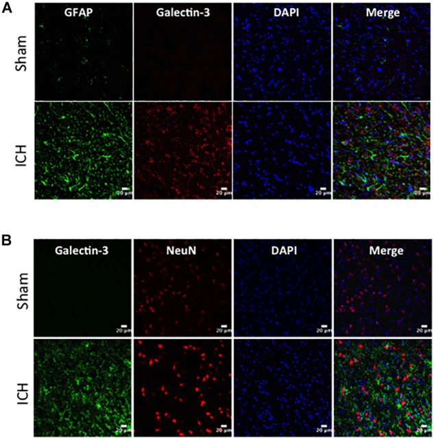
Cellular localization of Galectin-3 after ICH. Brain sections were double immunostained with (A) Galectin-3 and GFAP, and (B) Galectin-3 and NeuN. Galectin-3 expression was absent in either GFAP-positive or NeuN-positive cells. Scale bar = 20 μm; n = 3 per group.