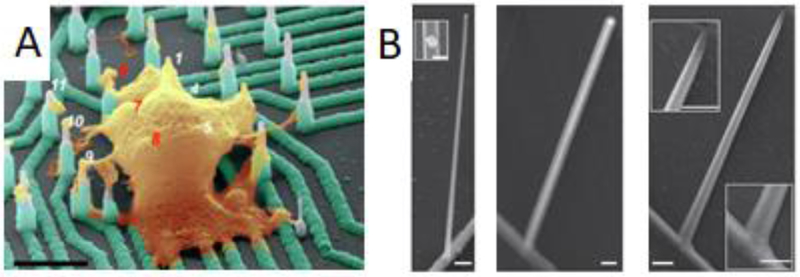
Figure 2.
Vertical structures for electrophysiology. A) Doped silicon nanoelectrodes record intracellular action potentials from primary and stem cell-derived neurons. Scale bar: 4 um. (R. Liu et al. 2017) B) Nanotube channels increase field effect transistor performance. Left: germanium branch on silicon nanowire. Inset gold nanodot on nanowire. Middle: structure after coating with aluminum oxide. Right: Hollow nanotube forms the transistor channel after etching of germanium core. Scale bars: 200 nm for all but left inset (100 nm). (Duan et al. 2011)

