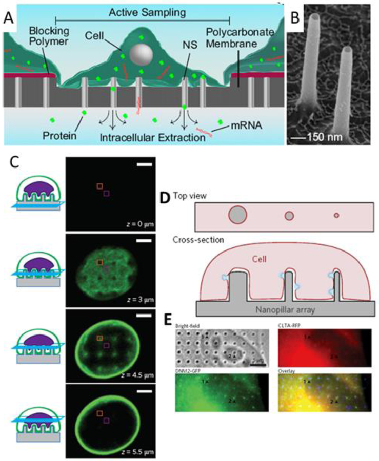
Figure 5.
Cellular assays. A) Schematic of nanostraw sampling of intracellular molecular contents. (Cao et al. 2017) B) SEM of alumina nanostraws. (Cao et al. 2017) C) Confocal z-stack of images depicting nanopillar-induced nuclear deformation. The nuclear envelope is labeled with GFP-Sun2. Scale bars 3 um. (Hanson et al. 2015) D) Schematic of Nanopillar-induced curvature for studies of endocytosis. E) Collocalized immunostaining for clathrin and dynamin2 allows correlation between degree of curvature and protein accumulation. (Zhao et al. 2017)

