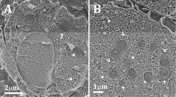
Figure 3:
(A, B): Cryo SEM micrographs of high pressure frozen and freeze fractured macrophage cells after 48 h incubation with 50 μg/ml acLDL. The samples were etched for 5–20 min at −105°C in order to expose the outer surface features and enhance the contrast between the cytoplasm and the unetched lipid objects. The cells are filled with objects (indicated by arrows) that have the typical texture of lipid-rich particles, most probably lysosomes and/or lipid droplets.

