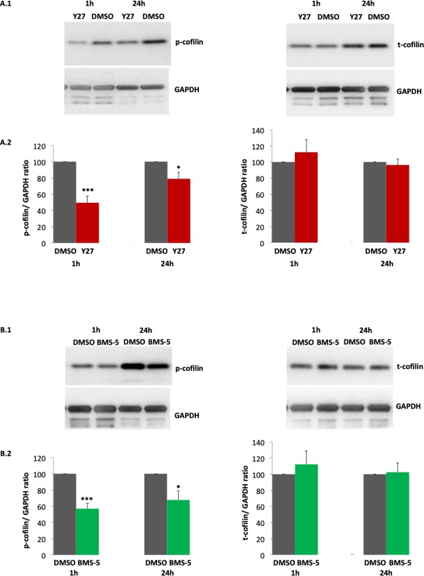
Figure 2.

ROCK or LIMK inhibition, by 30 μM Y727632 (Y27) or 30 μM BMS-5 respectively, decreases phosphorylation of cofilin in porcine retinal explants 1 and 24 hours after detachment. Comparison between ROCK or LIMK inhibition and control was conducted with detached retinal explants from the same eyes. DMSO was the control condition (A.1, B.1). Antibodies against p-cofilin and t-cofilin labeled 19-kDa bands. GAPDH, from the same SDS-PAGE gel, served as an internal loading control. (A.2, B.2) Quantification of cofilin bands, normalized by GAPDH. Note, there was no change in t-cofilin with drug treatment. (A) n = 4 animals, 4 retinal explants per group; (B) n = 4 animals, 4 retinal explants per group; Student's t-test, *P < 0.05, ***P < 0.001.
