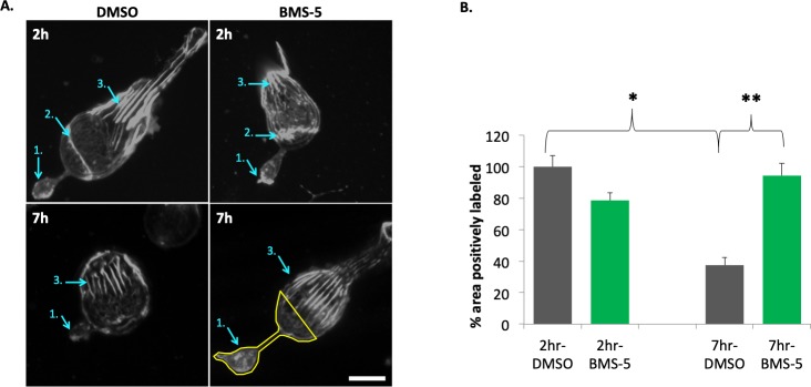
Figure 6.
LIMK inhibition (10 μM BMS-5) stabilizes actin filaments in the basal (axonal) region of rod photoreceptors after injury. (A) Examples of actin filament staining with phalloidin in rod photoreceptors, 2 and 7 hours in control (DMSO) and treated (BMS-5) cultures. Cell in 2 hour-control has an outer segment. Arrows: 1, axon terminal; 2, zonula adherens; 3, calycal processes. Lower right panel indicates the ROI (outlined in yellow) used for quantification, which includes the terminal, axon if present, and the basal-most portion of the nuclear pole to which the axon terminal retracts. Comparison of treated and control cells at 2 hours and 7 hours shows that the actin filaments in the calycal processes remained structurally intact in the presence or absence of an outer segment. (B) Quantification of fluorescent signal for F-actin in ROI of rod cells at 2 and 7 hours in culture. Percentage of total ROI area with positive labeling for F-actin was measured and normalized (control group set at 100%). In control, a significant loss of staining at 7 hours suggests depolymerization of actin filaments has occurred. BMS-5 prevents this loss. Optical sections, 1 μm. Scale bar: 10 μm. n = 3 animals, 12 culture dishes, 101 rod cells; 1-way ANOVA with post hoc Tukey's test, *P < 0.05, **P < 0.01.

