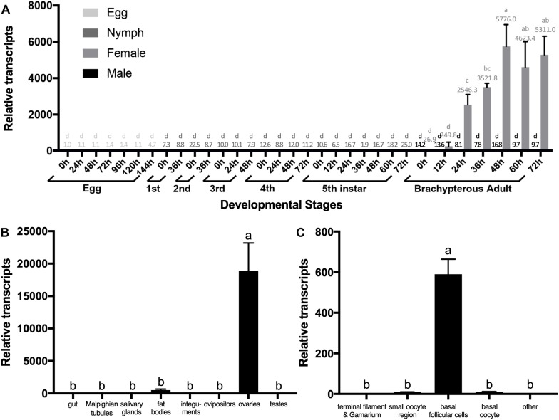
FIGURE 2.
The expression pattern of NlESMuc. (A) NlESMuc expression according to BPH developmental stage. Samples were collected from eggs, nymphs, and brachypterous male and female adults. The NlESMuc mRNA level in eggs at 0 h was designated 1 and relative expression levels for each sample were marked in different colors according to the color code. (B) NlESMuc expression in BPH tissues. The digestive tract (n = 50), Malpighian tubes (n = 50), salivary glands (n = 80), fat bodies (n = 50), ovipositors (n = 30) and integument (n = 20) of brachypterous females were dissected 48–120 h after emergence, while the inner reproductive organs (n = 30), testes (male), and ovaries (female) of brachypterous adults were dissected 48–120 h after emergence. The NlESMuc mRNA level in the integument was designated 1. (C) NlESMuc expression in BPH ovary. The terminal filament–germarium region, small oocyte region (containing small developing oocytes < 800 μm and active follicular cells), basal oocytes (oocytes ∼850 μm in length), basal follicular cells (follicular cells around the basal oocytes), and other parts (oviduct, bursa copulatrix, spermatheca, pouched gland) were dissected from brachypterous adult females 48–120 h after emergence. The NlESMuc mRNA level in the other parts was designated 1. Nl18S and RPS11 were used as internal control genes. Data represent the relative NlESMuc mRNA levels. Data are the mean ± SEM from three independent experiments. Columns followed by a common letter are not significantly different by the HSD-test at the 5% level of significance in each histogram.

