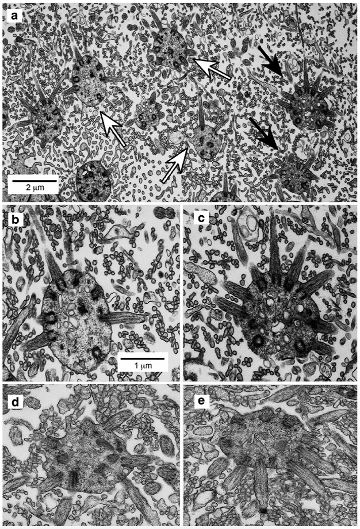
Fig. 7.

Immunoelectron microscopy showing the localization of GFP-labeled dendritic knobs and cilia of olfactory sensory neurons in tangential sections at the surface of the epithelium. A. Dendritic knobs appear as spherical profiles that contain multiple basal bodies from which the cilia extend. Both unlabeled knobs (e.g., white arrows) and GFP− labeled knobs (black arrows) extend cilia characterized by the presence of microtubules and basal bodies. Also present in the image are microvilli of sustantacular cells, appearing as smaller circular profiles lacking defined cytoplasmic structure or details. B. & C. Higher magnifications of corresponding unlabeled (leftmost white arrow) and GFP-labeled (upper black arrow) dendritic knobs in panel A. Scale bar in panel B also applies to panel C. D & E. Examples of dendritic knobs from control tissues not exposed to primary antiserum. No dark knobs are evident.
