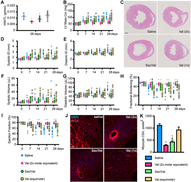
Figure 1: SAC/VAL prevents cardiac hypertrophy and loss of function associated with pressure overload-induced heart failure more effectively than equimolar valsartan.
A. Heart weight/tibia length ratio reveals a decrease in pressure overload-induced cardiac hypertrophy with SAC/VAL, which significantly reduces hypertrophy relative to saline or equimolar valsartan alone. n = 10, 10, 12, 14. One-way ANOVA with Tukey’s multiple comparisons test. B. LV mass by echo confirms the significant effect of SAC/VAL relative to equimolar valsartan. For A, B, D-I: n = 10, 10, 12, 14. Two-way ANOVA with repeated measures (time). * = p < 0.05 compared to saline; & = p < 0.05 for SAC/VAL compared to equimolar valsartan. C. H&E staining of transverse sections through the papillary muscle further confirms SAC/VAL’s significant effect against hypertrophic remodeling in response to pressure overload. Scale bar = 1mm. D-E. Systolic/diastolic LV internal diameter (ID) increase is prevented by SAC/VAL but not the molar equivalent of valsartan. F-G. Systolic/diastolic volume increase is prevented by SAC/VAL but not the molar equivalent of valsartan. Two-way ANOVA with repeated measures (time). H. Fractional shortening is significantly improved by SAC/VAL, but not the molar equivalent of valsartan. Two-way ANOVA with repeated measures (time). I. Ejection fraction is significantly improved by SAC/VAL but not the molar equivalent of valsartan. Two-way ANOVA with repeated measures (time). J. Myocyte cross sectional area (CSA) is visualized by wheat germ agglutinin (WGA) staining. Scale bar = 100 μm. K. CSA is increased due to concentric hypertrophic remodeling induced by pressure overload (saline), and is decreased in SAC/VAL-treated mice significantly more so than equimolar valsartan. One-way ANOVA, Tukey’s multiple comparisons test. n = 5, 5, 6, 7. * = p < 0.05 compared to saline; & = p < 0.05 for SAC/VAL compared to equimolar valsartan.

