Figure 8:
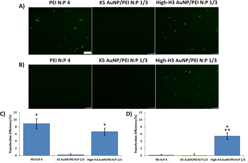
CHO-K1 transfection with GFP-encoding pDNA. Representative fluorescence microscopy images of GFP expression 24 h post-transfection with the indicated hybrid AuNP complexes or PEI polyplexes either (A) without or (B) with heparin. Complexes were formed at an overall N:P ratio of 4, with an N:P = 1 contribution from the AuNPs and a N:P = 3 contribution from the PEI. Quantification of transfection efficiency (C) without or (D) with heparin using flow cytometry. All results are shown as the mean ± standard deviation of data collected from three independent experiments. * indicates a significant difference from zero (p < 0.05). ** indicates a significant difference from PEI polyplexes. Scale bar = 250 μm.

