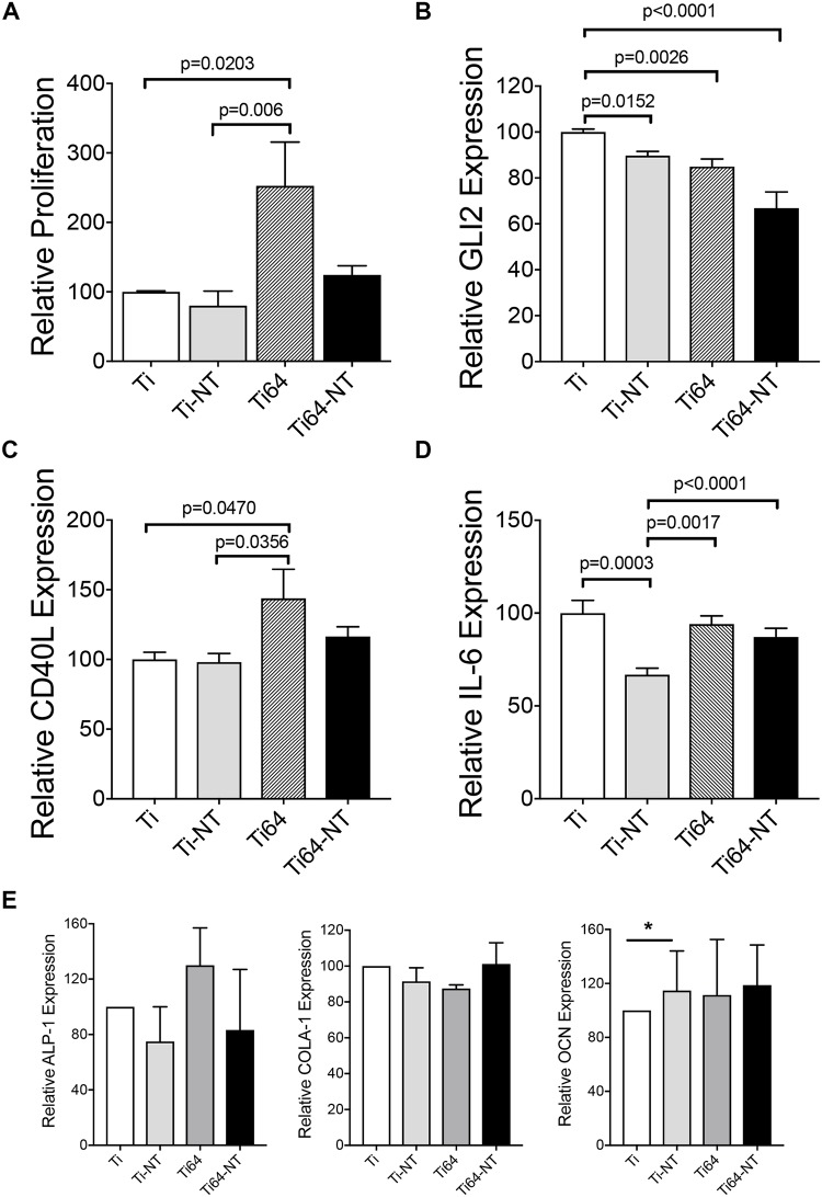
Fig 6. Effect of composition and surface morphology on growth of BMSCs and inflammation.
a) Saka cells were allowed to adhere on material as indicated in methods for 3 days followed by XTT assay to determine cell proliferation. qRT-PCR for the inflammatory markers b) GLI2, c) CD40L and d) IL-6. E) Saka cells were allowed to adhere A similar experiment was performed to determine the expression of differentiation markers alkaline phosphatase 1 (ALP-1), Collagen A-1 (COLA-1) and osteocalcin (OCN) on the different substrates by qRT-PCR. Data are presented as averages of 2 independent experiments, each performed in triplicate and the bars represent means ± SEM.

