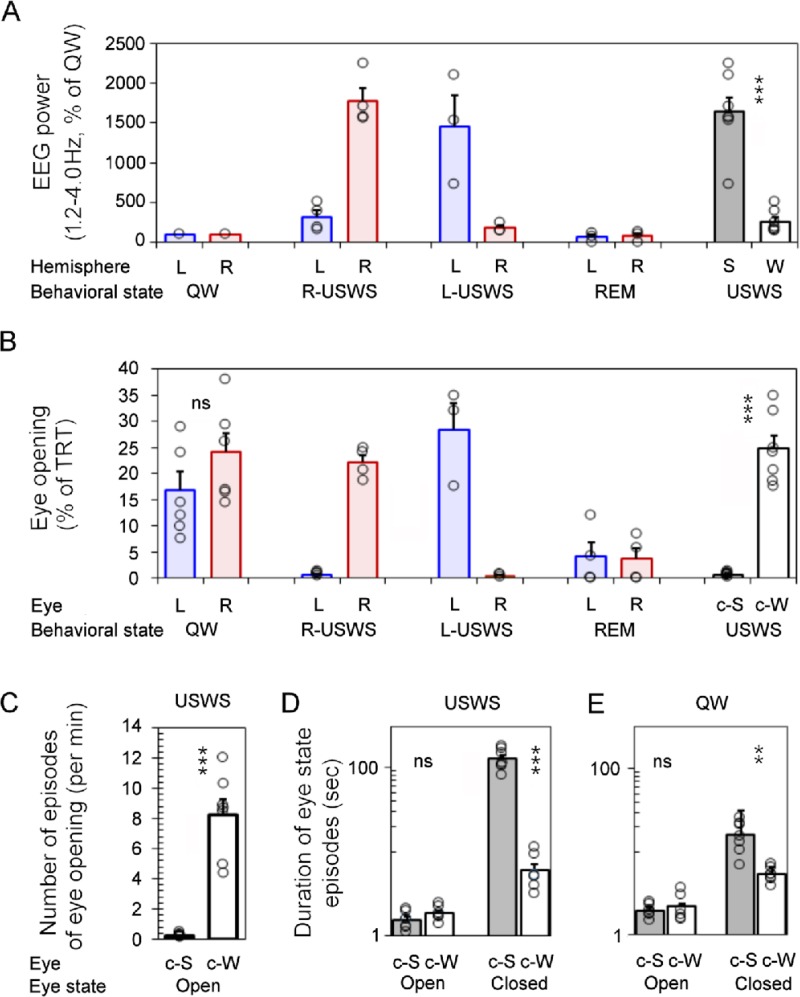
Fig 3. Association between behavioral state and eye state in fur seals in water.
(A) Standardized EEG power in the range of 1.2–4.0 Hz in two cerebral hemispheres (shown as percent of the average power in QW in the corresponding hemisphere) during QW, R-USWS, L-USWS, USWS (R-USWS and L-USWS) and REM sleep in 2 fur seals. (B) Total amount of the open state of the left and right eye during waking and sleep states. (C-E) Number and duration of the episodes of eye opening and closure during USWS (C, D) and QW (E). S and W correspond to sleeping and waking hemispheres. L and R correspond to left and right hemispheres or eyes. c-S and c-W correspond to which eye is contralateral to the sleeping and waking hemisphere, respectively. During QW the eyes were classified as c-S or c-W based on the adjacent USWS. Open circles represent the means for individual USWS, QW and REM sleep episodes recorded in 2 seals (S2 Table). Bars are grand average (mean) ± SEM for all episodes of QW and USWS. The difference between the power and the state of the eye was tested for all 7 episodes of USWS and 7 adjusted episodes of QW recorded in seals A and B. **,*** Statistical significance (the paired T-test) with p<0.01 and <0.001, respectively. ns–difference was not significant (p>0.05; S1 File).

