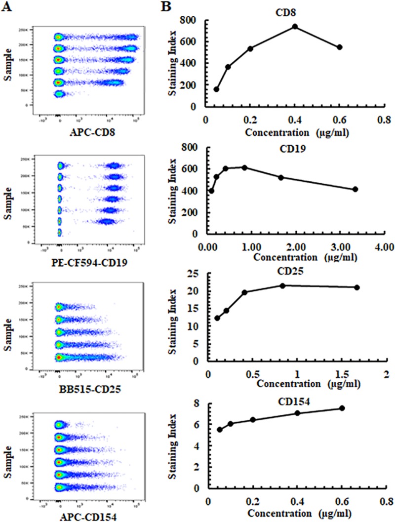
Fig 2. Four representative fluorochrome-conjugated antibody titrations.

(A) Concatenated pseudocolor data at 5–6 antibody concentrations for APC-CD8 (RPA-T8), PE-CF594-CD19 (HIB19), BB515-CD25 (2A3) and APC-CD154 (TRAP1). (B) Staining indexes and antibody concentrations for APC-CD8, PE-CF594-CD19, BB515-CD25 and APC-CD154. APC-CD8, PE-CF594-CD19, BB515-CD25 were assessed on lymphocytes (excluding CD4+ cells), lymphocytes or CD3+CD4+ T cells respectively. APC-CD154 was evaluated on CD3+CD4+ T cells of PBMCs that were activated with CD3/CD28 beads overnight. The final antibody concentrations used in the study for APC-CD8, PE-CF594-CD19, BB515-CD25 and APC-154 were 0.05 μg/ml, 0.38 μg/ml, 0.83 μg/ml, 0.3 μg/ml respectively.
