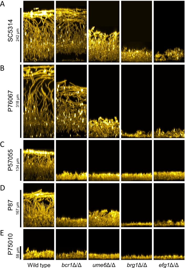
Fig 1. Biofilm side-view projections.
Wild-type and mutant strains in each clinical isolate background were assayed for biofilm formation under in vitro conditions. All strains were grown on silicone squares in RPMI + 10% serum at 37°C for 24 hours. Fixed biofilms were stained using Concanavalin A, Alexa Fluor 594 conjugate, then imaged by confocal microscopy. Representative sections from each biofilm are shown; relevant genotypes are given beneath each column. Scale bars indicate the depth of the corresponding wild-type biofilm. Strain backgrounds: A. SC5314. B. P76067. C. P57055. D. P87. E. P75010.

