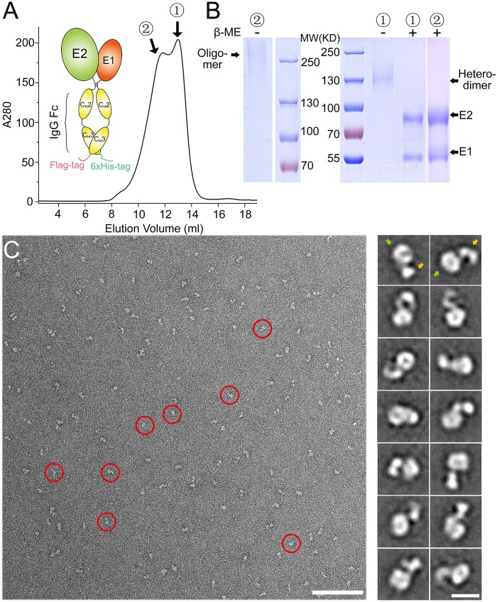
Fig 1. Expression of HCV E1E2 as an Fc-tagged heterodimer in insect cells.
(A) Schematic representation of the Fc-tagged HCV E1E2 heterodimer and the SEC profile of the purified E1E2-Fc. The heterodimeric and oligomeric peaks of E1E2-Fc are labeled as 1 and 2, respectively. (B) SDS-PAGE of the purified E1E2-Fc under reducing and non-reducing conditions for the two peaks shown in (A). (C) A negative staining EM image showing the heterodimeric E1E2-Fc particles (left; red circles; bar, 100 nm). The representative 2D averaging classes are also shown (right; bar, 10 nm). The head and the tail regions are indicated by green and orange arrows, respectively.

