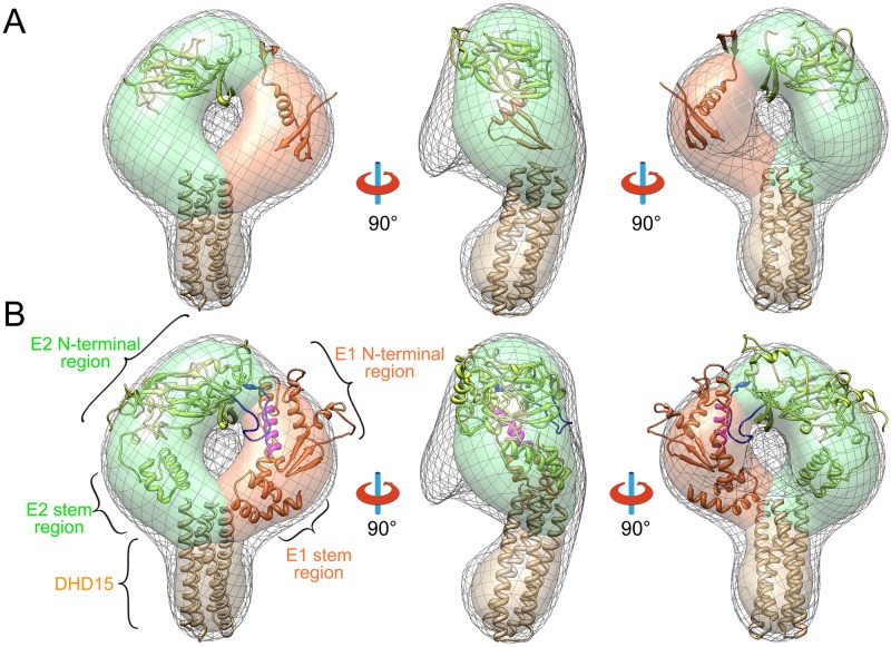
Fig 4. Three-dimensional EM reconstruction and the structural model of HCV E1E2 heterodimer.
(A) Three views of the 3D reconstruction of E1E2-DHD15 (low-density contour: gray mesh; high-density contour: surface). The solved structures of E2 (PDB entry: 4MWF, green), E1 (PDB entry: 4UOI, orange) and DHD15 (brown) are put into the EM density showing that a large portion of E1 and E2 are missing in the known crystal structures. The densities corresponding to E2, E1 and DHD15 are colored in green, orange and brown in the high-density contour, respectively. (B) Three views of the coevolution based E1E2 structural model fitted into the EM reconstruction. The hyper variable region 2 (HVR2, blue) of E2 and the putative fusion peptide (magenta) of E1 are shown in the structural model.

