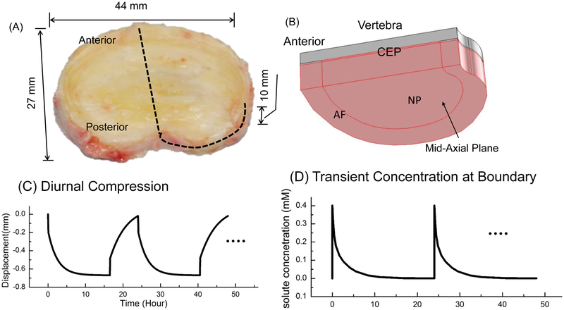
Figure 1:
(A) Picture of a human lumbar intervertebral disc (IVD, L2–3, non-degenerated). (B) Schematic of a quarter of the IVD-vertebra segment used in the FEM analysis (due to symmetry). (C) Schematic of the diurnal compression (on a quarter of the disc). (D) Schematic of the transient antibiotic concentration at the disc boundary. NP: Nucleus pulposus. AF: Annulus fibrosus. CEP: Cartilaginous endplate.

