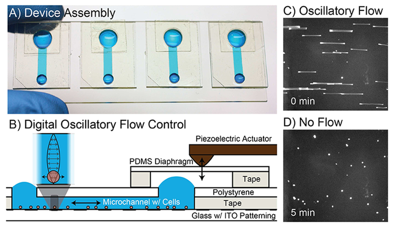
FIG. 1.

Microfluidic interface. A) Each ITO-pattemed glass slide contains four microfluidic devices to facilitate sample loading and control of adhesion for sensing. Blue food coloring is used for illustration. B) Devices are constructed on the ITO-patterned glass using razor-printed double-sided adhesive, polystyrene, and PDMS. C) unlabeled cells are imaged in bright-field mode with an extended exposure to show the oscillatory motion of each cell as a streak. D) The same unlabeled cells are imaged in bright-field mode after stopping flow to allow the cells to interact with the antibody functionalized surface.
