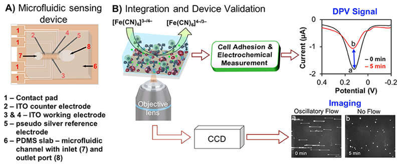
SCHEME 1.

A) Schematic drawing of a fully assembled microfluidic sensing device. B) Schematic of electron transfer reaction at the working electrode surface in the absence (curve a) and in the presence of melanoma cells (curve b) and simultaneous microscopic image acquisition of cell adhesion during oscillatory flow (image a) and after stopping flow (image b). [Note: color in figures and images are for illustration only].
