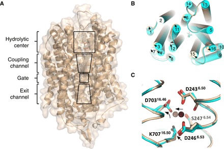
Fig. 1. Overview of the TmPPase structure.

(A) Monomer showing the location of the hydrolytic center, coupling channel, ion gate, and exit channel. (B) Top view of the superposition of the TmPPase:IDP:ATC (wheat) and TmPPase:IDP complex (cyan) structure showing relative TMH movements (arrow) upon binding of ATC. (C) Superposition of the gate region between two structures [TmPPase:IDP:ATC (wheat) and TmPPase:IDP complex (cyan)]. D2466.53, D70316.46, and Na+ slightly moving away (arrow) relative to their positions in the TmPPase:IDP structure. Violet-purple and pink spheres are for Na+ of TmPPase:IDP and TmPPase:IDP:ATC, respectively.
