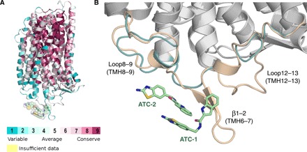
Fig. 6. The conservation of ATC binding site among mPPases.

(A) Structure of TmPPase colored according to the sequence conservation among the set of 16 pathogenic mPPases. The pale green surface indicates the location of the ATC binding site. (B) Comparison of the ATC binding sites of the crystal structure of TmPPase (wheat) and a homology model of PfPPase (teal). Carbon atoms of ATC are presented as pale green sticks. Blue, nitrogen; red, oxygen; yellow, sulfur.
