
Fig. 1. Myocardial PDE1A expression in mouse hearts overexpressing CryABR120G or wild-type CryAB.
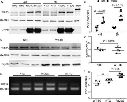
Total protein and total RNA from ventricular myocardial samples were used for Western blot analyses (A to D) and RT-PCR (E and F), respectively. GAPDH, glyceraldehyde-3-phosphate dehydrogenase. (A and B) Representative images (A) and pooled densitometry data (B) of Western blot analyses for PDE1A in 4-month-old (4M) or 6-month-old (6M) CryABR120G tg (R120G) and their NTG littermate mice. For each time point, four NTG (two males and two females) and four R120G (two males and two females) mice were used. (C and D) Western blot analysis for myocardial PDE1A in CryABWT tg (WT-TG) and NTG control mice at 6 months. Littermate NTG (one male and two females) and WT-TG (two males and two females) were used. (E and F) Representative image (E) and pooled densitometry data (F) of RT-PCR analyses for myocardial PDE1A mRNA levels in NTG, R120G, and WT-TG mice at 6 months. For each genotype, two males and two females were tested. P values are derived from two-tailed unpaired t test with Welch’s correction (B and D) or one-way analysis of variance (ANOVA) followed by Tukey’s multiple comparison tests (F). ns, not significant.