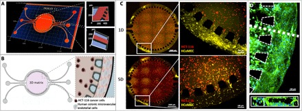
Fig. 1. Design and characterization of the microfluidic chip.

(A) Profilometer characterization: 3D map of the microfluidic chip. Representative image of quality control and characterization of the microfluidic chip features. The central chamber is 5 mm in diameter and 126 μm in depth, with a separate inlet and outlet for hydrogel injection. The two lateral channels, each 100 μm in width and 126 μm in depth, are not interconnected, opening up the possibility of perfusing two distinct solutions. (B) Schematic of chip design and zoom-in image of the concept of colorectal tumor-on-a-chip model: Round microfluidic central chamber in which HCT-116 cancer cells are embedded in Matrigel; HCoMECs are seeded in the side channels to form a 3D vessel-like assembly. (C) Establishment of a microvascular 3D microenvironment of colorectal tumor-on-a-chip: HCT-116 CRC cells embedded in Matrigel (in central chamber, stained in red) and HCoMECs (in lateral microchannels, stained in yellow) at day 1 (1D) and day 5 (5D) of culture. Endothelial cells start to invade the central chamber filled with Matrigel in response to VEGF presence. (D) Fluorescence microscopy image of microfluidic lateral channel mimicking prevascularization with HCoMECs after 5 days of culture [4′,6-diamidino-2-phenylindole (DAPI) blue, nuclei; phalloidin green, F-actin filaments]. Representative fluorescence microscopy close-up image of microchannel cross section showing endothelial cells aligning and creating an endothelialized lumen within the microchannel.
