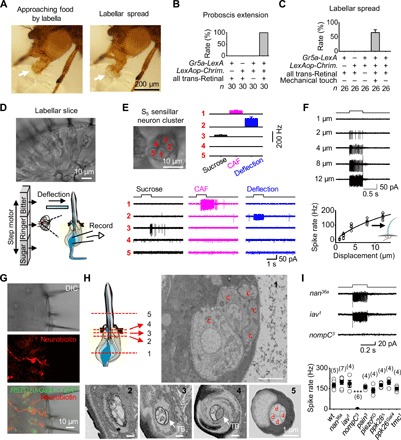
Fig. 1. Labellar spread and mechanical detection by taste bristles.

(A) Labellar spread during natural feeding. The extended labella approach the food with two labellar lobes closed (left, arrow), followed by opening of the two labellar lobes upon food contact (right, arrow). (B) Sweet gustation does not drive labellar spread. Optogenetic activation of the CsChrimson-expressing Gr5a-GRNs produces proboscis extension but does not elicit labellar spread. Light stimulation: 1 s, 617 nm. (C) Mechanical stimulation drives labellar spread. Optogenetic activation of Gr5a-GRNs elicits proboscis extension; labellar lobes spread apart upon touching a coverslip. Light stimulation: 1 s, 617 nm. (D) A labellar slice preparation. Top: Differential interference contrast (DIC) image of a labellar slice. Bottom: Gustatory stimulation (left); mechanical stimulation and single-cell suction-pipette recordings on a sensory neuron (right). (E) Sensory responses of the S5 sensillar neurons. Top: DIC image of five neurons in one S5 sensillum (left); collective data of spike responses of the five neurons averaged across different S5 sensilla, in which all five neurons are recorded successfully one by one (right; n = 6). Bottom: Spike responses of individual neurons in one S5 sensillum to mechanical deflection (10 μm) and gustatory stimuli [50 mM caffeine (CAF) and 100 mM sucrose]. (F) Deflection dependence of mechanosensory responses. Top: Spike responses to sensillar deflection. Bottom: Collective data (n = 8). The fit is with a Boltzmann function. (G) Bristle MSNs do not extend dendrites into the bristle cavity. Top: DIC image of a labellar slice. Middle: Neurobiotin labeling of a mechanosensitive neuron. Bottom: Overlay of the DIC, green fluorescent protein (GFP), and neurobiotin images. GMR57C10-Gal4 is a pan-neuron driver. (H) Structure of sensory neurons in a bristle by electron microscopy. Top: Illustration of five positions (left); the five neuronal cell bodies of the same bristle at position 1 (right). Bottom: Five dendrites of the bristle at position 2; four dendrites and one tubular body at position 3; the large tubular body at position 4; four dendrites and no tubular body in one sensillar cavity at position 5. c, cell body; TB, tubular body; d, dendrite. (I) NOMPC-dependent mechanosensitivity. Top: Representative spike responses to sensillar deflection. Bottom: Collective data of firing rates (calculated as five times the total spikes during the 200-ms deflection). Mechanical deflection, 200 ms and 10 μm. ***P < 0.001. wt, wild-type.
