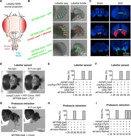
Fig. 4. Bristle MSNs and peg MSNs control distinct feeding behaviors.

(A) Schematic illustration of axonal projections in SEZ by labellar MSNs. (B) Peg (left) and bristle (right) slices. NP7506-Gal4 labels all peg MSNs and two to four bristle MSNs (top); nompC-LexA labels all peg MSNs and bristle MSNs (middle); nompC-LexA, LexAop-FRT-CsChrimson.mVenus-FRT together with NP7506-Gal4/UAS-FLP label exclusively bristle MSNs (bottom). (C) Distinct axonal projections of peg MSNs (top), peg and bristle MSNs (middle), and bristle MSNs (bottom). Red, tdtomato; green, GFP; blue, anti-nc82 immunostaining. (D) Bristle MSNs drive labellar spread. Optogenetic activation of bristle MSNs drives labellar spread. Light stimulation: 1 s, 617 nm. (E) Collective data of labellar spread by bristle MSNs. (F) Labellar spread does not require peg MSNs. (G) Peg MSNs drive proboscis retraction. Optogenetic activation of peg MSNs triggers proboscis retraction. Light stimulation: 1 s, 617 nm. (H) Collective data of proboscis retraction by peg MSNs. (I) Proboscis retraction requires peg MSNs.
