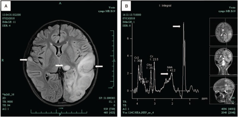
Figure 1.

Brain MRI of patient showed the left temporal, occipital and parietal lobe, cortex and subcortical of right temporal lobe, and left thalamus had abnormal signals (A) MRS showed NAA peak in the lesion area decreased significantly, and raised lactate peak in the lesion at 1.3 ppm (B). MRI = magnetic resonance imaging, MRS = magnetic resonance spectroscopy, NAA = N-acetyl aspartate.
