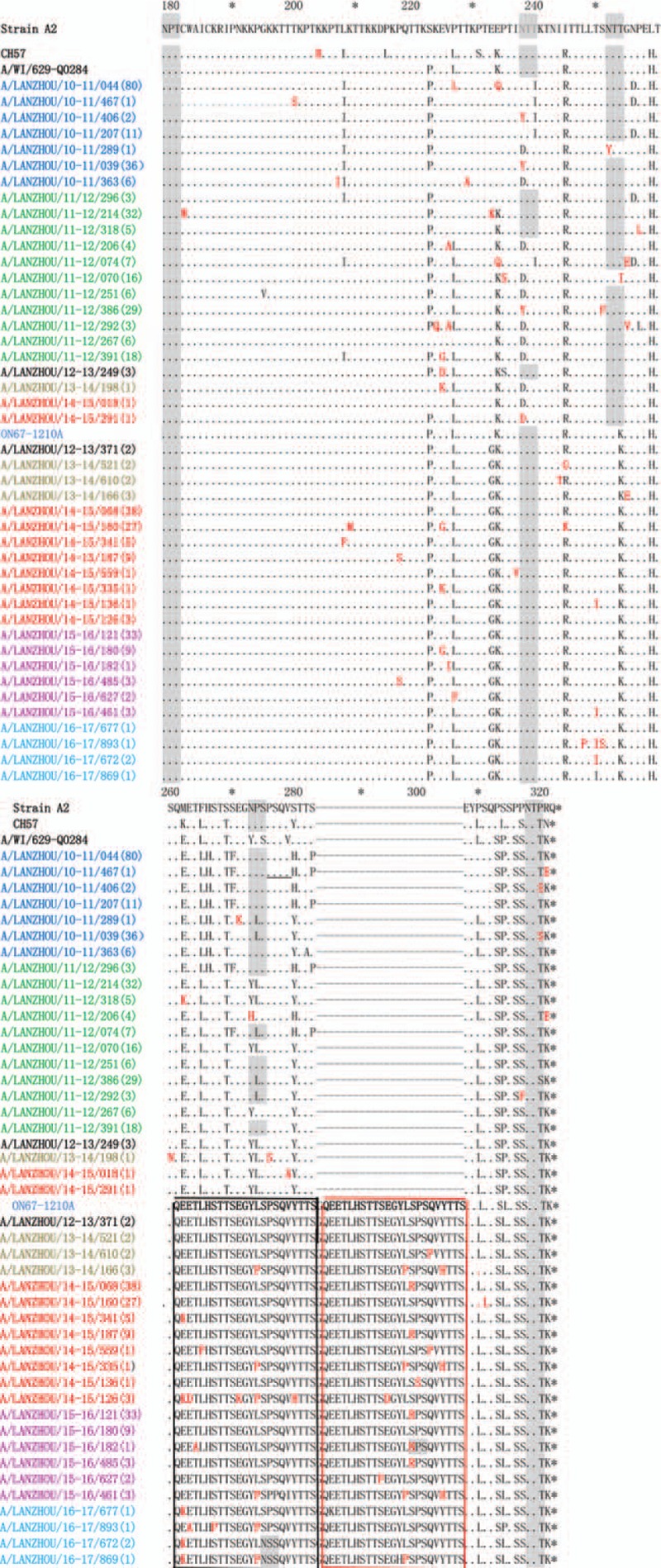
Figure 3.

Alignment of deduced amino acid sequence of the G protein of RSV-A strains isolated in Lanzhou during the 2010 to 2017 epidemic seasons. Alignments are shown relative to the sequences of prototype strain A2, genotype GA2 strain (CH57), NA1 strain (A/WI/629-Q0284), and ON1 strain (ON67-1210A). The AAs shown correspond to positions 179 to 298 of the second hypervariable region of RSV-A strain A2 G protein. Numbers in parentheses indicate the total number of identical strains. Dots indicate nucleotides identical to the strain A2; the sequence of the ON1 prototype is shown only for clarity. Dashes indicate the gap corresponding to the nucleotide insertions; asterisks indicate stop codons. Black and red boxes indicate the duplicated region (homologous portion and insertion); mutations are shown in red and bold. Predicted N-glycosylation sites are shaded in gray. Color of sequence names indicates the epidemic season—blue: 2010/2011; green: 2011/2012; black: 2012/2013; brown: 2013/2014; red: 2014/2015; purple: 2015/2016; turquoise blue: 2016/2017.
