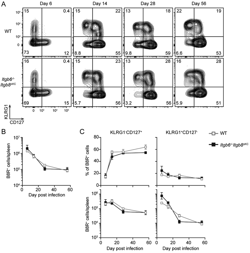
Figure 3. Intravenous vaccinia virus (VV)-induced CD8+ T cells are maintained normally in Itgb6−/−Itgb8ΔKC mice.
Splenic cells at the indicated time following intravenous VV infection in WT or Itgb6−/−Itgb8ΔKC mice were analyzed for KLRG1−CD127+ memory precursors and KLRG1+CD127− terminal effectors by flow cytometry. (A) Representative plots gated on B8R+ cells are shown. (B) Total number of B8R+ cells, and (C) frequency (top panels) and total number (bottom panels) of KLRG1−CD127+ or KLRG1+CD127− B8R+ cells are shown. Data are representative of two separate experiments with at least four mice per each time point. Data are means ± s.e.m.

