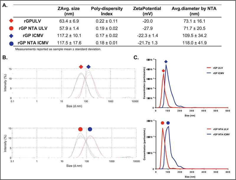
Figure 3. Size distributions of rGP ULV and ICMV formulations.

(A) Table of intensity-based nanoparticle sizes, and polydispersity indices and zeta potentials from Zetasizer measurements, along with number-based nanoparticle diameters by nanoparticle tracking analysis. Measurements reported as mean ± standard deviation. (B) Intensity-based and (C) number-based size distributions of antigen-loaded ICMV formulations, compared with the respective ULVs.
