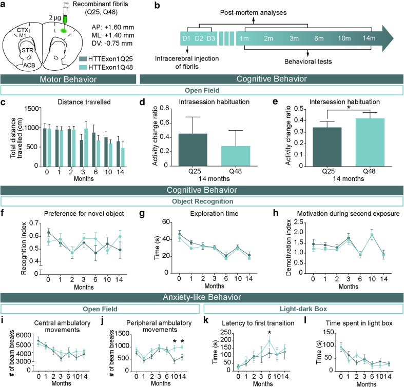
Fig. 3.
Manifestation of behavioral impairments in adult WT mice injected with HTTExon1Q48 fibrils. Atlas coordinates for intracerebral injections, treatment legend (a) and experimental timeline (b). Animals underwent motor, cognitive and anxiety-like behavioral tests from 1 to 14 months post-injection. Motor behavior was assessed using total distance traveled in 60 min in the open field (c). At 14 months post-injection, short-term memory was evaluated by assessing the change in distance traveled between the first 5 and the last 5 min of testing (d) and long-term memory was assessed by calculating the change in distance traveled in the first 5 min of baseline to 14 months post-injection (e). Cognitive performance was assessed at each time point by measuring the preference for the novel object (f). The absence of confounding factors was determined by measuring the exploration time (g) and motivation during the exposure to the novel object (h). Anxiety-like behavior was assessed in the open field by quantifying the distance traveled in the center of the open field (i) and in the periphery (j) as well as in the light–dark box by latency to leave the dark box (k) and total time spent in the light box (l). Data are expressed as mean ± SEM. Baseline HTTExon1Q25 n = 12, HTTExon1Q48 n = 12; 1 month HTTExon1Q25 n = 11, HTTExon1Q48 n = 11; 2 months HTTExon1Q25 n = 10, HTTExon1Q48 n = 10; 3–14 months HTTExon1Q25 n = 9, HTTExon1Q48 n = 9. Statistical analysis was performed using a students’ unpaired t test for individual time points and a two-way ANOVA followed by Tukey’s post hoc test for across time graphs. *p < 0.05. ACB nucleus accumbens, AP antero-posterior, CTX M1 primary motor cortex, D day, DV dorso-ventral, m month, ML medio-lateral, STR striatum

