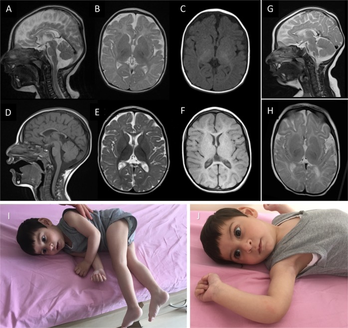
Fig. 1. MRI findings and phenotypes of our patients with ASNSD.
Brain MRI of patient 1 obtained at 4 months old (upper row, a–c) and 3.5 years old (lower row, d–f). Delayed myelination is best appreciated on the T1-weighted axial image obtained at 4 months old (c) and on the T2-weighted axial images obtained at 3.5 years old (e). The thin corpus callosum is displayed well in both sagittal T2- and T1-weighted images (a+d), while cerebral atrophy and the simplified gyration prominent in the frontal lobes were found on follow-up imaging (d–f). Brain MRI of patient 2 obtained at 4 months of age (g+h). The sagittal T2-weighted image (g) shows a very thin corpus callosum. Delayed myelination is better appreciated in image (h). The clinical phenotype of patient 2 (i+j) included absent fixation, reduced attention to external stimuli, and central hypotonia, which made him unable to sit without support

