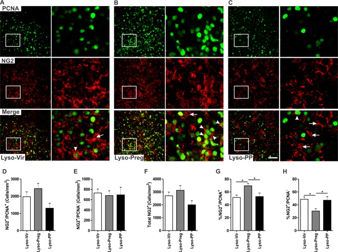
Figure 2.
Division of OPCs is enhanced in the vicinity of the corpus callosum 7 days following lysolecithin-induced demyelination in pregnant rats. (A–C) are immunofluorescent staining images of PCNA (green) and NG2 (red) in lysolecithin-injected corpora callosa of virgin, pregnant, and postpartum animals respectively. (D) The bar graph shows that there was no significant difference in the cell density of dividing OPCs (NG2+/PCNA+, arrowheads), non-dividing OPCs (NG2+/PCNA−, arrows) (E), or the total number of NG2+ cells (F) between the three experimental groups (Lyso-Vir: n = 6, Lyso-Preg: n = 5, Lyso-PP: n = 4, p > 0.05). (G) The bar graph shows that focally demyelinated corpus callosa of pregnant animals have a significantly higher percentage of NG2+/PCNA+ cells when compared to those seen in either virgin or postpartum animals (p < 0.05). (H) The bar graph shows that demyelinated corpus callosa of pregnant animals had a significantly lower percentage of NG2+/PCNA− cells compared to those seen in either virgin or postpartum animals (p < 0.05). Data are presented as mean ± SEM. Scale bar = 50 µm.

