Figure 3.
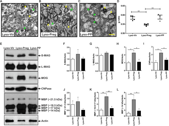
Axonal myelination is increased 7 days post-lysolecithin-induced demyelination in the corpus callosum of pregnant rats. (A–C) show representative TEM images of the corpus callosum post-lysolecithin injection. Green arrows indicate unmyelinated axons while yellow arrowheads indicate the myelinated ones. (D) Graph shows comparison of the g-ratio between the three experimental groups. The axons seen in the corpus callosum of pregnant rats had a significantly lower g-ratio when compared to those seen in virgin and postpartum animals (Lyso-Vir: n = 4, Lyso-Preg: n = 4, Lyso-PP: n = 3, p < 0.01). (E) Representative figures of western blot for the different myelin proteins. No significant difference was detected in the expression of S-MAG (F) or L-MAG (G) in demyelinated corpora callosa of the three experimental groups (Lyso-Vir: n = 6, Lyso-Preg: n = 5, Lyso-PP: n = 4, p > 0.05). The expression of both MOG (H) and CNPase (I) were significantly higher in the pregnant rats when compared to those seen in either virgin or postpartum rats (p < 0.05). The expression of the 21.5 kDa MBP isoform (J) was not different between the three experimental groups (p > 0.05). Pregnant rats had a significantly higher expression of 18.5 + 17 kDa isoforms (K) when compared to either virgin or postpartum rats (p < 0.01). The expression of the 14 kDa MBP isoform (L) was significantly increased in pregnant rats when compared to either virgin (p < 0.01) or postpartum (p < 0.05) rats. Results are presented as mean ± SEM. Scale bar = 1 µm.

