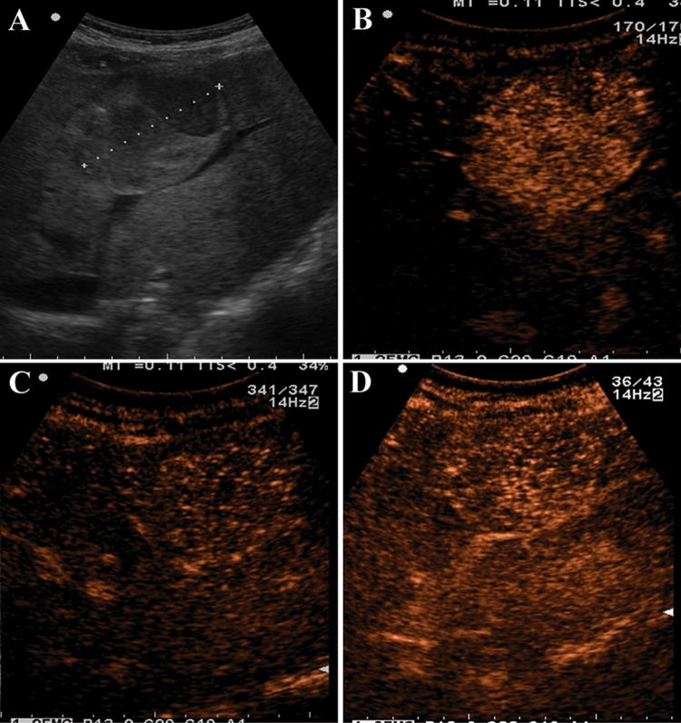
Fig. 2.
A 29-year-old woman who had glycogen storage disease underwent ultrasound for raised liver function tests (no previous oral contraceptives intake). a Grayscale ultrasound showing a single nodule of 6 cm with a mixed echogenicity in the right lobe of the liver (white arrow). b Contrast-enhanced ultrasound showed a rapid enhancement and centripetal filling pattern in the arterial phase (21 s after SonoVue injection). c Contrast-enhanced ultrasound in the portal phase showed no washout (50 s after SonoVue injection). d In the late venous phase the nodule is still iso/hyper-enhanced as compared to the surrounding liver parenchyma (≈ 2 min after SonoVue injection)

