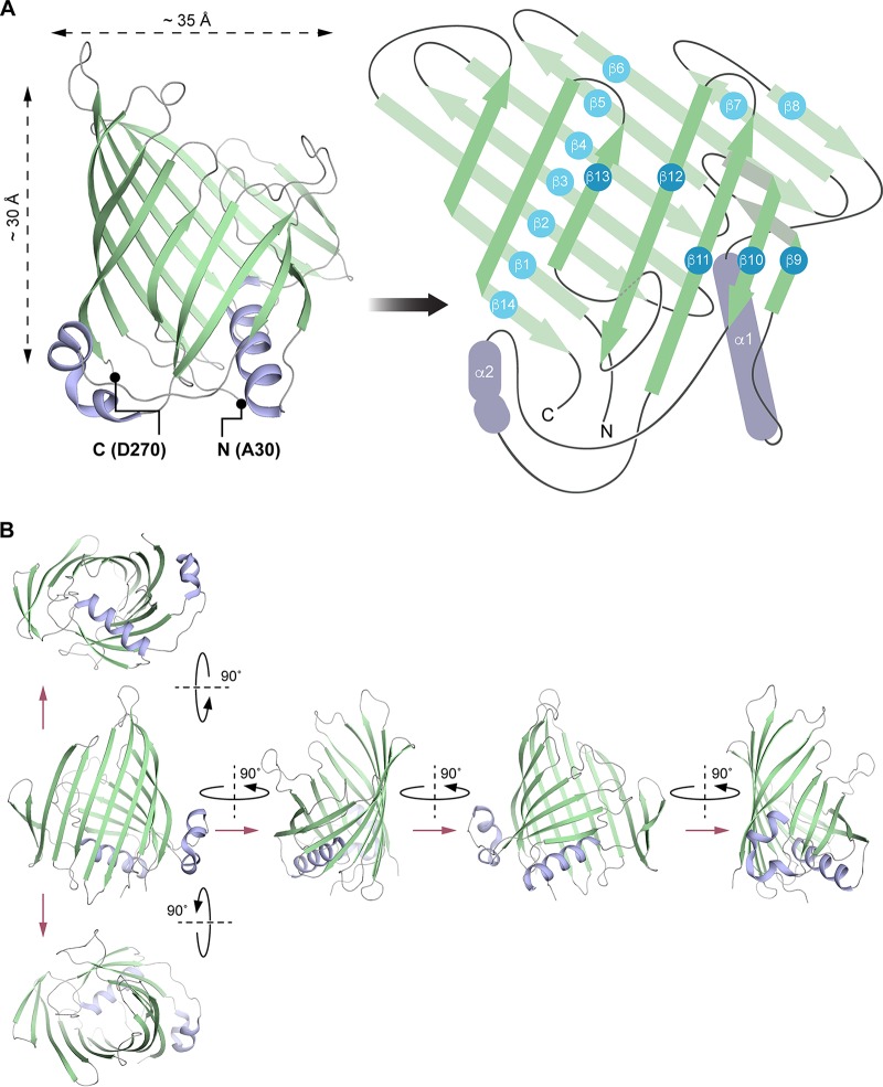
FIG 5.
EipB adopts a β-spiral fold. (A, left) X-ray structure of EipB. EipB consist of 14 β-strands (in green) and 2 α-helices (in violet). The N terminus (A30) and the C terminus (D270) are reported on this structure. (Right) Simplified representation of EipB. The color code is the same as for the left panel. (B) Different orientations of EipB structure. The color code is the same as for panel A.

